Objective: We aimed to evaluate the angiographic findings and outcomes of bronchial artery embolization in tuberculosis patients and to compare them with those of non-tuberculosis patients.
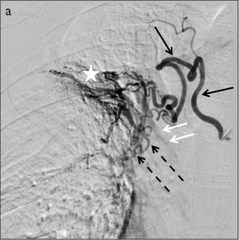
Materials and Methods: Patients who underwent bronchial artery embolization in a single interventional radiology department with hemoptysis were reviewed. A total of 89 patients (66 males and 23 females; mean age 52.71±15.37) were incorporated in the study. The patients were divided into two groups: tuberculosis group (n=36) and non-tuberculosis group (16 malignancy, 22 bronchiectasis, 6 pulmonary infection, 5 chronic obstructive pulmonary disease, 4 idiopathic; n=53). Angiography and embolization procedure were performed by interventional radiologists with 5, 10, and 20 years of experience. Angiographic findings were classified as tortuosity, hypertrophy, hypervascularity, aneurysm, bronchopulmonary shunt, extravasation, and normal bronchial artery. Chi-square test was used to compare angiographic findings between tuberculosis and non-tuberculosis patient groups.
Results: Bronchopulmonary shunt was found to be significantly higher in the tuberculosis group as compared to that in the non-tuberculosis group (p=0.002). Neither of the groups showed a statistically significant difference with respect to recurrence (p=0.436).
Conclusion: Bronchial artery embolization is a useful and effective treatment method of hemoptysis in tuberculosis. Evaluation of bronchopulmonary shunts in patients with tuberculosis is critical for the reduction of catastrophic complications.
Cite this article as: Sarıoğlu O, Capar AE, Yavuz MY, et al. Angiographic Findings and Outcomes of Bronchial Artery Embolization in Patients with Pulmonary Tuberculosis. Eurasian J Med 2020; 52(2): 126-31.

.png)
_In%20Progress-1%20(1)_page-0001.jpg)

.png)
.png)
